In this section

Images in this gallery were taken on the Nikon D-Eclipse C1 Confocal Microscope by students who conducted independent research on campus. 

GnRH-3-GFP (green) and active capase-3 (red) expression in a 36 hpf Danio rerio embryo.
Noah Dillon ’20: Zebrafish Embryo

2019 Image Contest Entry - GnRH-3-GFP (green) and active capase-3 (red) expression in a 36 hpf Danio rerio embryo. Epifluorescence microscopy was used to capture images of green fluorescent protein and Alexa Flour 594 at 100x. Images were merged together using ImageJ software.

YFP fluorescence in a whole leaf sample of Nicotiana benthamiana epidermal cells.
Meghan Bacher ’17: N. benthamiana epidermal cells

2016 Image Contest Entry - YFP fluorescence in a whole leaf sample of Nicotiana benthamiana epidermal cells. Individual cells are shaped like puzzle pieces. Fluorescence indicates the interaction of ACS5 and 14-3-3 ω, which serves as a positive control for Bimolecular Fluorescence Complementation (BiFC) experiments conducted with FBS1 and other 14-3-3 proteins.


Drosophila melanogaster wing disc dissected from wandering larvae.
Bridget Alexander ’18: Fruit Fly Wing Disc

2016 Image Contest Entry - Drosophila melanogaster wing disc dissected from wandering larvae. The wing disc was stained with DAPI (blue) and antibodies (red) to identify the presence of dPRL. GFP (green) was found to be present. Identifying the presence of dPRL was used as a checkpoint before any crossing of lines could occur.


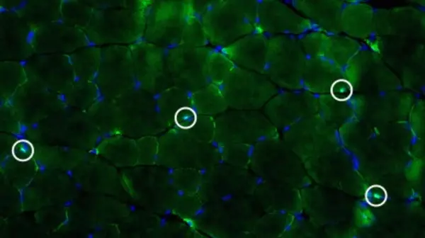
Satellite cell identification using immunofluorescence in mouse plantaris muscle.
Mike Pang ’16: Mouse Muscle

2015 Image Contest Entry - Satellite cell identification using immunofluorescence in mouse plantaris muscle. 10 μm muscle cryosections were immunolabeled with anti-Pax7 primary antibody (green) and stained with DAPI (blue). Satellite cells were quantified by counting cells that had co-localized Pax7 and DAPI signal, shown as circled cells in all panels.


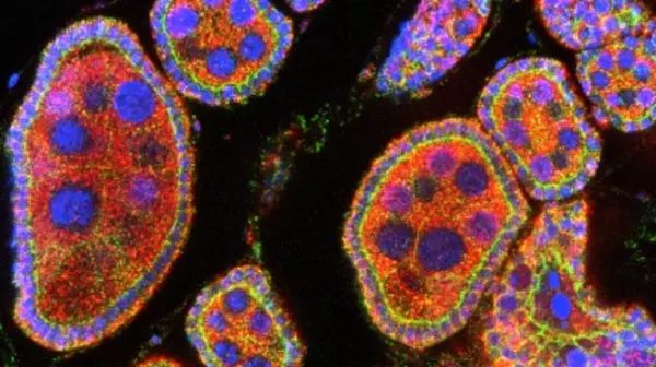
Drosophila melanogaster egg chambers in the ovary.
Kara Sherva ’15: Fruit Fly Egg Chambers

2015 Image Contest Winner - Drosophila melanogaster egg chambers in the ovary. Stained for DNA (blue), the membrane-bound cell adhesion molecule E-cadherin (green), and the gene product of the potential oncogene dPRL-1 (red). dPRL-1 is seen in the cytoplasm of the larger nurse cells and in the membranes of the nurse and follicle cells, co-localizing with E-cadherin.


Filamentous bacterial mat samples from the high sulfide sediment of Commencement Bay were prepared by Fluorescence in situ Hybridization (FISH).
Cheyenne Dewey ’16: Bacterial Mat

2014 Image Contest Winner - After identification by 16S rRNA gene sequencing, filamentous bacterial mat samples from the high sulfide sediment of Commencement Bay were prepared by Fluorescence in situ Hybridization (FISH). The microbial community in the mat was then visualized using epifluorescence. The genera Arcobacter (green) and Sulfurovum (red) were labeled by the fluorophores FITC and Cy3, respectively. Environmental DNA is stained with DAPI (blue).


Microplastic particle found in a Mytilus mussel collected along Ruston way in Tacoma, WA.
Nick Lyon ’16: Microplastic

2014 Image Contest Entry - Microplastic particle found in a Mytilus mussel collected along Ruston way in Tacoma, WA. Particles of this chemical composition auto-fluoresce at near-UV light. This is some of the first proof that plastics discarded by people can end up in the bodies of organisms previously thought to be largely unaffected by that variety of pollution. 


Root nodules of Scotch broom (Cytisus scoparius) prepared as cross-sections then illuminated with confocal microscopy.
Michaela Alden ’13: Root Nodules

2013 Image Contest Entry - Root nodules of Scotch broom (Cytisus scoparius) prepared as cross-sections then illuminated with confocal microscopy. The plant tissue auto-fluoresces in response to three wavelengths of light, revealing what may be vascular bundles around the inner edge of each nodule. 

Less-than-24-hour-old Drosophila melanogaster embryo, stained for DNA (DAPI) and dPRL-1(Cy3).
John Valenzuela ’13: Fruit Fly Embryo

2013 Image Contest Entry - Less-than-24-hour-old Drosophila melanogaster embryo, stained for DNA (DAPI) and dPRL-1(Cy3). dPRL-1 (fuchsia-color) is our protein of interest in Professor Leslie Saucedo’s lab, as it is overexpressed in many mammalian cancers.